Size Does Matter: X-Ray Image Scaling Devices
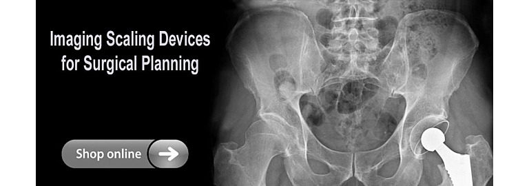
It is well known that x-rays used for surgical planning vary in magnification. This variation exists in part because of the diversity of techniques used by x-ray technologists, patient positioning, and the patient’s size. Even with strict imaging protocols, the magnification of an x-ray image can vary. A thin patient lying on an imaging receptor is only a few inches above the plate, giving little distance for the X-rays to diverge. In a larger patient, the distance may be twice as far from the plate, allowing the divergent beam to spread much more before it hits the plate. This can result in a magnification factor of approximately 20%. Scaling an x-ray image is mandatory to assure the highest level of accuracy throughout the planning process, especially orthopedic templating. Does size matter when it comes to magnification of an x-ray and surgical planning? Absolutely! Preoperative planning aims to reduce the risk of intraoperative fracture and decrease overall surgical time. Images that are used for surgical planning must be accurately scaled to allow for accurate implant selection.
Historically, orthopedic surgeons used precisely constructed plastic "overlays" matched up with anatomy in standard X-Ray films to calculate the magnification factor and, to a degree, determine the correct prosthetic to be used in surgery. The rapid introduction and implementation of digital x-ray rendered standard overlays useless. Soon orthopedic templating software packages became available for digital imaging, however, in order to effectively plan for surgery there was still a need to determine the magnification factor inherent in the image. Many different methods were used but all shared a degree of difficulty and often provided unacceptable results. What was needed was an x-ray marker with consistent dimensions from any angle that was capable of being positioned over a wide range of locations.
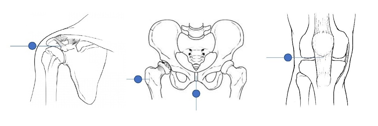
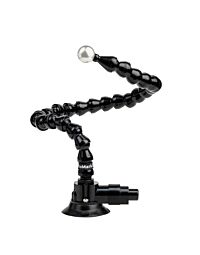
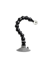
Scaling your x-ray begins with placement. The center of the x-ray scaling marker should be paced as closely as possible to the part of interest. Image magnification is measured to accurately scale the anatomy, by placing a calibration object or a radiopaque object of known dimension, in a precise position in the X-ray’s field of view. The main advantage to using spheres is that the spheres are entirely symmetrical. These calibration spheres are mounted on an adjustable flexible arm. The arm allows the object to be correctly positioned in the same plane as the anatomy of interest quite easily. By positioning the stainless steel calibration ball at the precise anatomical plane of the shoulder, hip, knee, etc., during x-ray imaging, surgical planning software is able automatically find the x-ray marker in the image, calculate the x-ray magnification factor, and determine the true size of the anatomy in question. This helps to ensure that the appropriate prosthetic is selected before surgery. External anatomy (e.g., the olecranon, ulnar styloid, medial malleolus) is often used to precisely position scaling markers as close to the region of interest as possible.
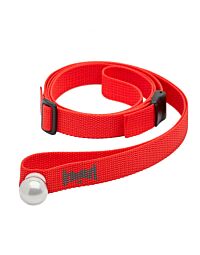
Z&Z Medical has a wide range of Image Scaling Devices in a variety of bases and attachment methods. A suction base allows for use on both horizontal table tops and on a vertical wall bucky making this stand a single source solution for all scaling needs. We have scaling devices with magnetic bases designed for use on ferrous surfaces, and clamp on models. These marking devices come with a flexible arm which can be easily extended at any time. and have a stainless-steel calibration ball which is solid and doesn't degrade when surrounded by larger body mass.
X-Ray Image Scaling Device Markers can also be used in the Veterinary market where orthopedic surgical templating software packages are used. For more information, visit www.zzmedical.com for all your imaging accessory needs.

