National Pet Cancer Awareness Month
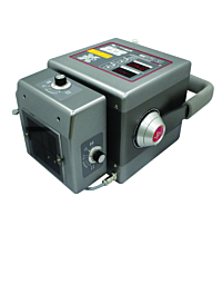
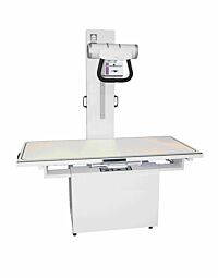
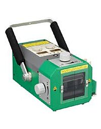
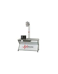
Both traditional and special contrast X-ray techniques are used to look for tumors in a pet's lungs, gastrointestinal tract, bladder and other internal organs. X-rays are usually used as the first imaging test to evaluate a pet's condition and determine whether the cancer has spread throughout the body.
CT is a specialized imaging technique available at referral centers or specialty hospitals. CT relies on the differences in density between tissues to form an image and the images of cross-sections of the body are generated by a computer. It is a superior technique compared to X-rays in evaluating cancer in the lung, the chest cavity and ribs, and is important for planning radiation therapy. CT scans can also be used to guide biopsies when a suspected mass needs further analysis.
Ultrasound refers to a technique used to examine internal organs in the abdomen and to also help guide biopsys. The veterinarian places a transducer emitting sound waves in contact with the area of interest (e.g. stomach), moves it around and views the structure of internal organs on a monitor in real time. It is routinely used to evaluate masses discovered during physical examination or to check for metastasis to liver, spleen or other organs. It is generally not used to evaluate structures that contain air such as the lungs since air prevents the transfer of sound waves.
MRI uses strong magnetic fields to create three-dimensional images created by a computer. MRI is extensively used to evaluate masses in the central nervous system such as the spine or brain and has been useful in providing images of soft tissues, joints, tendons, muscles and bone marrow.
Z&Z Medical understands the importance of Imaging animals and works closely with the Veterinary Field to provide a wide array of imaging accessories and supplies for both X-Ray, CT, Ultrasound and MRI for animal imaging. Visit our website to learn more or email info@zzmedical.com


